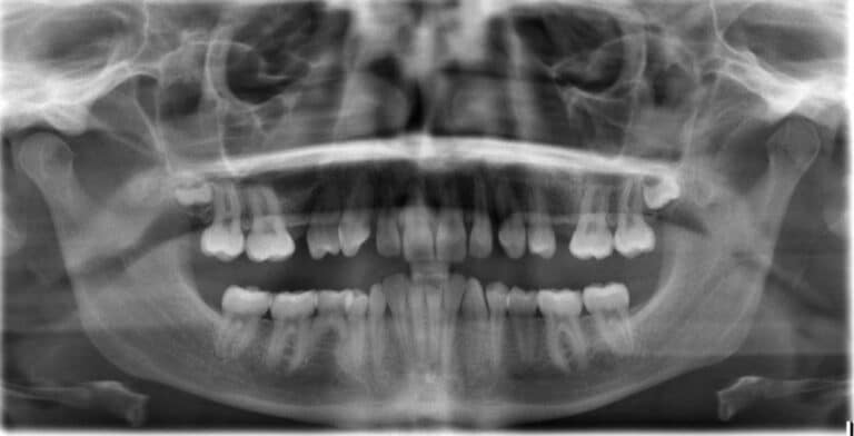
X-Ray – Panoramic Viewing
Basically, the panoramic x-ray is a standard part of orthodontic diagnosis. Not only is the jaw and all teeth with some neighboring regions be seen on this type of x-ray, but the maxillary sinuses are visible as well. Hidden cavities, inflammation of the jaw, and stored or displaced teeth are typical findings detected with this recording.
Jaw Joints
The temporomandibular joint is shown, but these findings are only to exclude very rough findings as deformations, fractures or tumors. An exact image diagnosis of the temporomandibular joint with the panoramic x-ray is not possible and usually not necessary.
Two Panoramic X-Rays per Treatment
The panoramic x-ray has a very good relationship between cost and benefits, which explains why we use it for initial diagnosis and following the needs of the case for continuing diagnosis.
Basically, most treatments require only two radiographs, with very few exceptions. Seldomly a panoramic x-ray is needed for final diagnosis. This is a considerable reduction in the widespread production of six or more cranial x-rays per orthodontic treatment.
Advantages of Digital X-Rays
Older panoramic x-ray equipment causes a radiation dose of 25-50 microsieverts, which is the considered as the normal background radiation at sea level of 5-10 days. In contrast, modern digital devices produce a radiation dose of approximately 20 microsieverts, which corresponds to the background radiation of four days. So relatively speaking, it is a small burden.